Cleft lip and palate and craniofacial syndromes

Cleft lip and palate

Cleft lip and palate (CLP) represents the most common congenital malformation of the face. Its incidence is estimated at 1 case per 500 to 1,000 births. Asiatic and aboriginal populations have a higher incidence compared to caucasians and African Americans.

This malformation affects both the lip and palate in 46% of cases. Thirty three percent involve the palate only and 21% are isolated cleft lips. CLP is more often unilateral (left side) and affects males more than females in a 2:1 ratio.

Treatment of CLP patients require a multidisciplinary team: pediatrician, speech pathologist, nutritionist, ENT surgeon, plastic surgeon, orthodontist and an oral and maxillofacial surgeon. In fact, kids with congenital malformation are at higher risk for respiratory, hearing, feeding and language problems if they are not appropriately managed.

Kids with CLP need several surgical procedures throughout their childhood. In the first months of life, the cleft lip is repaired (cheiloplasty). Thereafter, between the age of 6 months and 2 years, the repair of the cleft palate (palatoplasty) is done. At around 5 years of age the patient should be seen by an orthodontist to evaluate dental crowding as there is usually a deficit in vertical, antero-posterior and transverse growth of the upper jaw. Orthodontic appliances and braces are used to align the teeth and expand the palate as needed.

Different dental anomalies are associated with CLP: supranumerary teeth, dental malformation, and congenitally absent teeth (most commonly the lateral incisor). Supranumerary teeth are usually removed and absent teeth are replaced as indicated.

The oral and maxillofacial surgeon is also involved in alveolar bone grafting of the cleft. After palatal expansion, the surgeon will normally graft the cleft with the autogenous bone harvested from the anterior iliac crest.

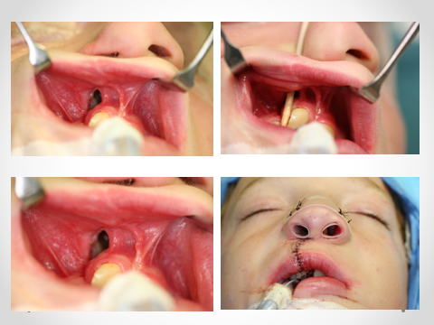
The alveolar bone graft at the cleft site will provide:

- Continuity of the alveolar bone of the upper jaw
- Bone support for the eruption of teeth adjacent to the cleft
- Bone support for possible future dental implants
- Closure of the oro-nasal fistula

The expertise of the oral and maxillofacial surgeon is called upon once again when patient’s growth has ceased and jaw corrective surgery is needed. Patients with CLP usually have a retrusive upper jaw; jaw corrective surgery is recommended is these cases. Moreover, the oral and maxillofacial surgeon will repair any residual oro-antral or oro-nasal fistula, graft deficient alveolar ridges, or perform cheilo-rhinoplasty.

Figure 1 Oronasal fistula cure, image courtesy of Dr. Valcourt*

Craniofacial syndromes

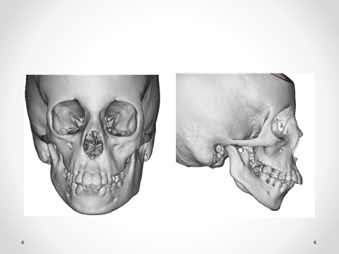
Hemifacial macrosomia is a deformity belonging to the spectrum of Goldenhar Syndrome (oculo-auriculo-vertebral dysplasia). It involves a unilateral hypoplasia of the face and affects mainly the middle and lower thirds.

This syndrome is associated with varying degrees of hypoplasia: the hypoplasia could lead to agenesis (complete absence) of the mandibular condyle as well as other parts of the lower jaw. The Goldenhar syndrome could also be associated with cleft lip and palate, hypoplasia or absence of different structures of the external and middle ear. The muscles and salivary glands on the same side also present varying degrees of hypoplasia. Goldenhar syndrome is confirmed when cardiac, cervical, and ocular anomalies are diagnosed.

Figure 2 Hemifacial microscopy, indicating the hypo-development of the right mandible. image courtesy of Dr. Valcourt*

The role of the oral and maxillofacial surgeon is to re-establish and improve the function and appearance of the maxillofacial complex. The hypoplastic mandibular condyle could be reconstructed with a costo-chondral graft followed eventually by corrective jaw surgery (Lefort I osteotomy and bilateral sagittal split osteotomies of the mandible) to align the jaws.

Multiple other syndromes can affect the maxillofacial region. These include Crouzon, Alpert, Pfeiffer, and Seathre-Chozen syndromes.

These syndromes are characterized by the presence of a cranial synostosis and alteration of the facial growth; premature fusion of the cranial suture will result in an abnormal cranial form. Lefort III osteotomies are needed to correct the cranial and frontal disharmony during childhood (between 7 and 10 years old).

These syndromes also involve the facial bones including the jaws. Therefore, some patients require orthognathic or corrective jaw surgery (Le Fort I osteotomy with or without bilateral sagittal split osteotomies of the mandible) once their growth is completed.

References :
1. Current Therapy In Oral and Maxillofacial Surgery, 1e. 1 edition. Philadelphia: Saunders; 2011. 1136 p.
2. Oral and Maxillofacial Pathology, 3rd Edition. 3rd edition. Saunders; 2008. 984 p.

*Annie-Claude Valcourt, Chirurgie buccale et maxillo-faciale, Hôpital de l’Enfant-Jésus, Québec

Sign In Mon Maxillo

Account details will be confirmed via email.

Reset Your Password